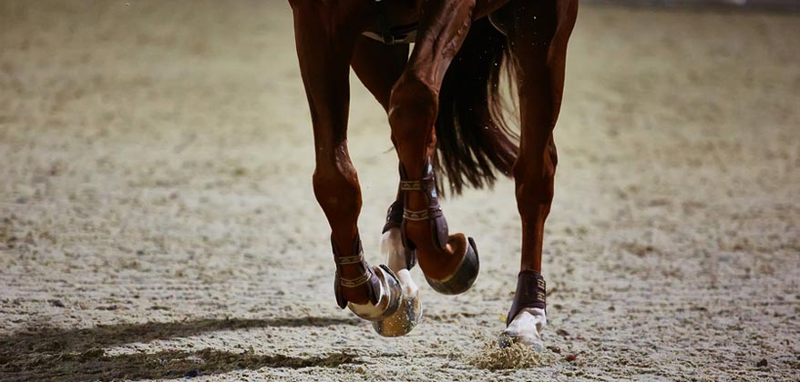
Your examination includes both static (standing still) and dynamic (moving) evaluations to determine if the horse is showing signs of pain associated with the musculoskeletal system. Static evaluation includes observation of symmetry, muscle mass (or atrophy), posture, and palpation for heat, pain, or swelling. Hoof testers are used to localize pain in the hoof. Dynamic evaluation includes observing the horse move at a walk and a trot in both the straight line and on the lunge circle.
Flexion tests are also preformed during a lameness exam to temporarily stress on a specific joint. If lameness is detected, diagnostic nerve blocks can be used to localize the source of the pain. Once pain is localized, diagnostic imaging including radiographs (x-rays) or ultrasound can be used to evaluate the soft tissue and boney structures in that area to determine the horse of the lameness.
Our veterinary surgeon responsible for your horse will discuss the potential benefits and risks associated with stem cell therapy. In most cases, we like to see the horse for follow-up assessment between one and three months after implantation.
If you have any specific questions about the procedure, costs, or the value of stem cells in treating injuries in horses, please contact Georgia Equine Veterinary Services and Hospital.
IRAP (Interleukin-1 Receptor Antagonist Protein): IRAP is ACS (autologous-conditioned serum) and is harvested from the horse’s own blood. IRAP is used to treat osteoarthritis and other inflammatory conditions in the joint. IRAP takes the horse’s own blood and processes. The serum is frozen and can be injected into the horse’s joint at anytime. From one blood pull on a horse we get about 10- 20mLs of IRAP serum to inject into joints. For osteoarthritis cases we recommend a series of IRAP to be injected once weekly for 3 weeks.
PRP (Platelet Rich Plasma): PRP is derived from the horse’s own blood. We pull blood from the horse and run it through our PRP machine and we get a solution of concentrated platelets which release growth factors that accelerate regeneration of injured tissues. PRP can be injected into tendon and ligament lesions. Recent research has been focused on injecting PRP into joints.
Purchase examinations may vary, depending on the intended use of the horse and the veterinarian who is doing the examination. Deciding exactly what should be included in the purchase examination requires good communication between you and your veterinarian. At GEVS we work with you to understand your expectations, and goals of the horse you are considering, establish costs involved and communicate with you or your agent to ensure no questions are unanswered.
GEVS offers pre-purchase examinations for prospective buyers. The pre-purchase examination includes a full physical exam (including ophthalmology, respiratory, musculoskeletal assessment), and a soundness exam. We offer radiograph packages and are happy to do any other diagnostics a client may want such as endoscopy, blood work(CBC and chemistry), or a drug screening at additional charges. If the prospective buyer cannot be present for the exam, we always communicate with him/her to discuss any findings and how he/she would like to proceed. We can email radiographs taken during the pre-purchase exam for the prospective buyer. We can perform pre-purchase exams here at the hospital or out on the road.
Our objective is neither to pass nor fail an animal; rather, it is to provide you with information regarding any existing medical problems and to discuss those problems with you so that you can make an informed purchase decision. We can advise you about the horse’s current physical condition, but we cannot predict the future. The decision to buy is yours alone to make. But we can be a valuable partner in the process of providing you with objective, health-related information.
Gastroscopy allows visualization of the esophagus and stomach to check for ulceration. Gastric ulcerations are erosions of the stomach lining due to prolonged exposure to acid produced by the stomach .Recent research reports that about 75% of competition horses have some grade of ulcers present. We have a 3 meter gastroscope that attaches to a full screen so both the doctor and client can view the horse’s stomach lining. Gastroscopy is a simple procedure, the fasted horse is lightly sedated and the scope is passed through the nasal passage down the esophagus into the stomach. Stomach ulcers or Equine Gastric Ulcer Syndrome (EGUS) is a common condition that affects many disciplines and breeds of horses. A study of dressage and hunters showed that as many as 60% were affected. Ulcers are common among all groups of horses that train or show.
There are factors that increase a horse’s risk of developing gastric ulcers: decreased grazing time or feeding limited amounts of hay, high grain diets, strenuous exercise, training, trailering, physical stress (from illness or other factors), and certain medications such as bute or banamine.
Symptoms of gastric ulcers are often difficult to detect, although they may have a serious effect on performance and horse comfort. Many horses will show only one symptom. Some example symptoms in adult horses include: attitude changes, dullness, poor or reduced performance, poor condition, decreased appetite, colic.
EGUS can be difficult to recognize since the symptoms may often be vague. The only accurate way to diagnose and determine the severity of ulcers is by gastroscopy.
The good news is that gastric ulcers can be successfully treated. Treatment involves the use of anti-ulcer medications as well as changes in management factors and feeding. There are also prevention protocols that can be designed for horses that participate in high risk activities.